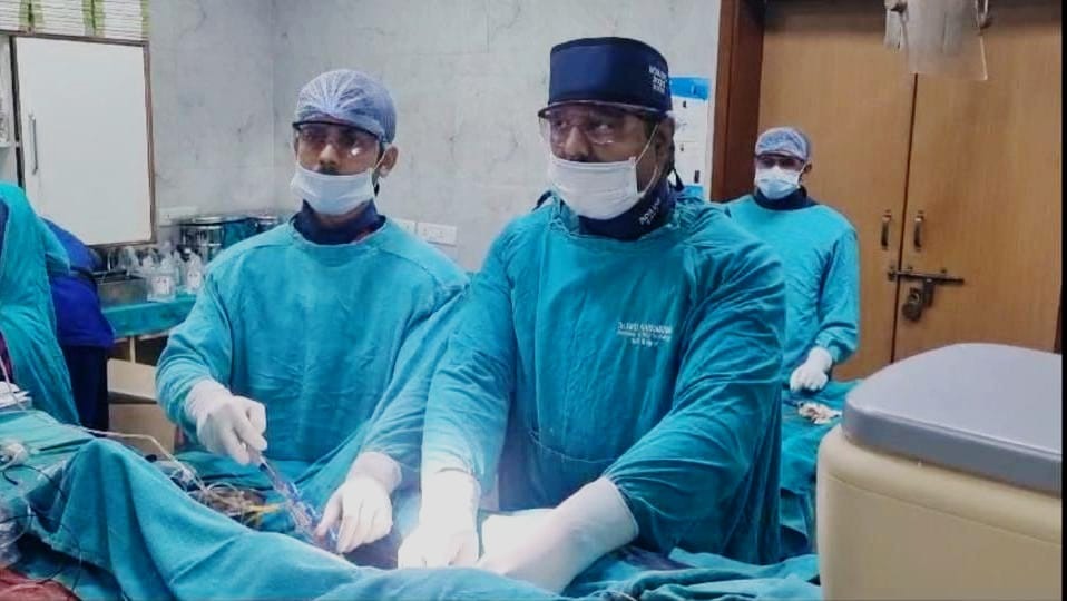
Raipur, July 3 (HS). At the Advanced Cardiac Institute (ACI) located at Dr. Bhimrao Ambedkar Memorial Hospital, a 66-year-old patient was successfully treated with excimer laser method for 100 percent blockage in the left artery supplying blood to the kidney and 90 percent blockage in the main vein of the heart.
According to the available medical literature, this is the first case in the world of treatment of complete block of kidney vein by laser angioplasty. In this treatment conducted under the leadership of Dr. Smit Srivastava, Head of Cardiology Department of ACI, the patient was saved from renal failure and heart failure by treating the patient's kidney artery i.e. renal artery and coronary artery simultaneously. These two interventional procedures are called Left Renal Artery Chronic Total Occlusion and In Stent Re-stenosis of Coronary Artery respectively.
In this case, for the first time, there was 100 percent occlusion of the kidney, due to which the BP was not coming under control. The kidney was getting damaged. If there was no timely treatment, the kidney would have failed. Giving information about the case, Dr. Smit Srivastava said that there was blockage in both the veins supplying blood to the patient's kidney. One had 100 percent blockage and one had 70-80 percent blockage. The main blockage was where the left renal artery starts. Due to this, the blood flow had stopped completely. Along with this, there was a blockage in the main vein of the patient's heart, for which he had a stent in a private hospital in 2023, which had stopped. This stent was completely blocked. Due to all these problems, the patient was suffering from heart failure, hypertension, difficulty in breathing and BP was not coming under control.
treatment done like this
First of all, the left renal artery which was 100% blocked, was opened using excimer laser due to a severe blockage. Then the path was widened using a balloon, which opened the tube completely by inserting a stent in it and normal flow was restored to the kidney. With the blockage being removed, changes in BP started coming and the BP decreased. By looking at the stent through intravascular ultrasound, it was confirmed whether it was properly placed in its place or not.
Due to previous angioplasty, more than 90 percent blockage was found inside the stent inserted in the left anterior descending artery, the main vein of the left side of the heart. This too was first opened by laser to open the blockage. Then the path was widened by ballooning and then the area of stent blockage was seen through intra vascular ultrasound. Since the blockage was inside the stent as well as outside the stent, it was decided to open that blockage by inserting a new stent. Both the blockages were treated by inserting an additional stent. The actual condition of the entire process was seen by doing IVUS. Ultimately both the procedures were successful. The patient is now fine and is ready to go home after being discharged.
 look news india
